


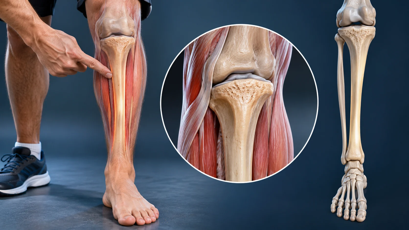
Where Is the Tibia Located in the Human Body?
The tibia is located in the lower leg, between the knee and ankle. Commonly called the shinbone, it is the larger and stronger of the two bones in the lower leg. The tibia runs along the inner side of the leg, while the fibula sits toward the outside. Together, these bones help support the body and enable everyday movements.
You can identify the tibia by gently feeling the hard ridge along the front of your shin. Unlike many other bones, part of the tibia lies close to the skin, making it easy to locate. Its upper end connects with the thighbone at the knee. The lower end helps form the ankle joint, allowing the foot to move.
Understanding where the tibia is located is important for learning about leg anatomy, bone injuries, and joint function. This bone plays a major role in standing, walking, running, and maintaining balance. Because it supports much of the body’s weight, it experiences considerable force during movement. Learning its basic structure can help explain common lower-leg conditions.
What Is the Tibia Bone?
The tibia is a long bone that forms the main weight-bearing structure of the lower leg. It extends from the knee to the ankle and works alongside the thinner fibula. The tibia is sometimes described as the shinbone because its front surface creates the prominent ridge of the shin. Its size and shape support both strength and movement.
The tibia consists of a long central shaft and two expanded ends. The upper end contributes to the knee joint, while the lower end participates in the ankle joint. Several muscles and ligaments attach to its surfaces. These connections help stabilize the leg and coordinate movements involving the knees, ankles, and feet.
Although the tibia is strong, it can still be injured through falls, accidents, and repeated physical stress. Fractures and overuse injuries may affect different sections of the bone. Understanding its structure helps explain why certain injuries cause pain in specific areas. Its anatomical features also provide important landmarks for medical examinations and imaging.
Tibia Location Compared With the Fibula
The tibia and fibula are the two long bones of the lower leg. The tibia sits on the medial, or inner, side, closer to the body’s midline. The fibula is positioned laterally, toward the outside of the leg. The tibia is considerably thicker because it carries most of the weight transmitted through the lower limb.
Both bones connect near their upper and lower ends through specialized joints and supporting tissues. A strong interosseous membrane extends between their shafts. This structure helps stabilize the relationship between the bones and provides attachment points for muscles. Although the fibula carries relatively little body weight, it contributes to ankle stability and muscle support.
A simple way to distinguish the bones is by feeling the front of your shin. The noticeable hard surface belongs to the tibia rather than the fibula. The fibula is more difficult to feel along much of its length because surrounding muscles cover it. Knowing this difference helps when interpreting descriptions of lower-leg pain or injuries.
Understanding the Anatomy of the Tibia
The anatomy of the tibia includes three major regions: the proximal end, the shaft, and the distal end. Each region has distinct structures that support different functions. The proximal end forms part of the knee, the shaft provides strength, and the distal end contributes to the ankle. These features make the tibia essential for lower-limb movement.
The tibial shaft has a roughly triangular cross-section, with several borders and surfaces. Its anterior border creates the noticeable shin ridge. The posterior surface contains anatomical markings associated with muscle attachments. The shaft also contains compact bone surrounding an internal cavity that houses bone marrow and contributes to the bone’s structure.
The upper and lower regions have expanded shapes that distribute forces across nearby joints. These ends contain smooth articular surfaces and important attachment points. Cartilage covers the relevant joint surfaces to support movement. Understanding these anatomical details helps explain how the tibia combines structural support with the flexibility needed for walking and other activities.
Proximal Tibia: The Upper Part Near the Knee
The proximal tibia is the upper portion of the shinbone, located directly beneath the knee joint. It is wider than the central shaft and includes two major regions called the medial and lateral condyles. Their upper surfaces form the tibial plateau. This area supports the lower end of the femur during standing and movement.
The tibial plateau is an important part of the knee because it receives substantial forces during weight-bearing activities. Cartilage and the menisci help distribute these forces and support smooth joint movement. Ligaments also contribute to knee stability through attachments near the upper tibia. Damage to this area can affect walking and other ordinary activities.
Another important feature is the tibial tuberosity, a raised area just below the front of the knee. The patellar ligament attaches here, connecting the kneecap to the tibia. This connection helps transmit forces produced by the quadriceps muscles. It is especially important during activities such as climbing stairs, standing from a chair, and extending the knee.
Tibial Shaft: The Long Middle Section
The tibial shaft forms the long central portion of the shinbone. It extends between the widened upper and lower ends and provides much of the bone’s structural strength. Its shape helps the lower leg tolerate forces during standing and movement. The shaft is also the region most people identify when referring to the shin.
The anterior border of the tibial shaft is particularly easy to feel beneath the skin. Because relatively little soft tissue covers this area, direct impacts can be painful. The surrounding surfaces provide attachment points for muscles involved in foot and ankle movements. These attachments help coordinate lower-leg function during everyday activities.
The tibial shaft can experience injuries ranging from bruising to fractures and stress-related bone problems. Repeated loading without sufficient adaptation may contribute to certain overuse conditions. The location and severity of symptoms depend on the affected tissues. Persistent pain along the shaft should be assessed rather than automatically attributed to ordinary muscle soreness.
Distal Tibia: The Lower Part Near the Ankle
The distal tibia is the lower end of the shinbone, where it contributes to the ankle joint. This region is wider than the central shaft and contains important joint surfaces. It connects with the talus, a bone located at the top of the foot. Together, these structures help support standing and controlled ankle movement.
A prominent feature of the distal tibia is the medial malleolus, the bony projection on the inner side of the ankle. You can usually feel this structure beneath the skin. It provides attachment points for ligaments that support ankle stability. The fibula forms the corresponding outer ankle projection, known as the lateral malleolus.
The distal tibia and fibula together form a socket around the talus, often called the ankle mortise. This arrangement allows the ankle to move while remaining stable under load. Injuries involving the distal tibia can affect joint alignment and mobility. Accurate medical evaluation is important when ankle trauma causes substantial pain, swelling, or difficulty bearing weight.
What Are the Main Functions of the Tibia?
The tibia performs several essential functions, beginning with supporting body weight. It transmits forces from the knee toward the ankle and foot during standing and walking. Its strong structure helps the lower limb remain stable under everyday loads. Without the tibia, normal upright movement would not be possible.
Another important function involves movement at the knee and ankle. The upper tibia participates in knee bending and straightening, while the lower portion helps form the ankle joint. Muscles and ligaments attached to the bone coordinate these activities. Together, these structures allow smooth transitions between standing, walking, climbing, and other movements.
The tibia also contributes to balance and lower-limb alignment. Its position helps maintain the relationship between the knee, ankle, and foot. This alignment influences how forces travel through the leg. Healthy bone and joint function allow the body to adapt to different surfaces and activities while reducing unnecessary stress on nearby tissues.
How the Tibia Connects to the Knee Joint
The knee joint connects the femur, tibia, and patella through a complex arrangement of cartilage, ligaments, and surrounding muscles. The upper surface of the tibia provides support for the femoral condyles. This relationship allows the knee to bend and straighten while supporting body weight. Joint stability depends on several structures working together.
The medial and lateral menisci sit between the femur and tibia. These fibrocartilage structures help distribute forces and contribute to joint stability. The anterior and posterior cruciate ligaments attach to the tibia and femur, limiting excessive movement. Additional ligaments along the sides provide further support during walking and other activities.
When the knee bends or straightens, the femur and tibia move relative to one another through coordinated rolling and sliding motions. Surrounding muscles control these movements and protect the joint. Injuries to the tibial plateau, ligaments, or menisci can affect normal knee function. Persistent instability, swelling, or significant pain requires medical assessment.
How the Tibia Supports the Ankle Joint
The tibia plays a central role in ankle stability because it forms the upper and inner portions of the main ankle joint. Its distal articular surface rests against the talus. This connection allows movements such as bringing the foot upward or pointing it downward. The fibula provides additional support along the outer side.
Strong ligaments stabilize the ankle and help control excessive motion. The medial malleolus provides attachment for the deltoid ligament complex, which supports the inner ankle. The distal tibia also connects with the fibula through the syndesmosis. This arrangement helps maintain appropriate alignment during standing, walking, and other weight-bearing activities.
Ankle injuries may involve bones, ligaments, or surrounding soft tissues. A fracture of the distal tibia can interfere with normal joint mechanics, particularly when the articular surface is affected. Accurate alignment is important for maintaining comfortable movement. Severe swelling, deformity, or inability to bear weight after an injury should receive prompt medical attention.
Common Tibia Injuries and Their Causes
Tibia injuries can occur through sudden trauma or repeated physical stress. Common examples include fractures, stress injuries, and problems involving nearby joints. Falls, sports collisions, and road accidents may cause acute fractures. Repetitive activities can also overload the bone when physical demands increase faster than the body can adapt.
Tibial fractures vary in location and severity. Some involve the shaft, while others affect the tibial plateau or ankle region. Symptoms may include localized pain, swelling, tenderness, and difficulty walking. Certain injuries can be serious even when external changes appear limited. Medical imaging is often necessary when a fracture is suspected.
Repeated loading may contribute to bone stress injuries, particularly in people involved in running or jumping activities. These problems may begin as localized discomfort that becomes more noticeable with activity. Continuing through persistent bone pain can worsen an injury. Early assessment helps distinguish bone stress injuries from muscle-related conditions and supports appropriate recovery planning.
Shin Splints: Pain Along the Tibia
Shin splints, also called medial tibial stress syndrome, are a common cause of exercise-related lower-leg pain. The condition typically causes discomfort along the inner border of the tibia. It often develops in people who perform repetitive weight-bearing activities. Runners and individuals beginning demanding training programs may experience symptoms when their loading tolerance is exceeded.
Pain from shin splints usually develops gradually rather than following one major injury. It may occur during exercise and sometimes continue afterward. Tenderness can affect a broader section of the inner shin. However, similar symptoms may occur with tibial stress fractures, making the exact pattern and severity important for diagnosis.
Management commonly involves adjusting aggravating activities and allowing symptoms to settle while maintaining appropriate movement. A qualified clinician may recommend progressive strengthening and a gradual return to activity. Persistent or sharply localized bone pain requires assessment for a possible stress injury. Ignoring worsening symptoms can delay recovery and increase the risk of more serious problems.
Tibial Fractures: Symptoms and Treatment
A tibial fracture occurs when part of the shinbone breaks because of excessive force or weakened bone structure. Fractures may develop after falls, accidents, or significant impacts. Some occur gradually through repeated stress. The symptoms and treatment depend on the fracture’s location, severity, and surrounding tissue involvement.
Common symptoms include pain, swelling, tenderness, and difficulty placing weight on the affected leg. More severe injuries may produce visible deformity or an inability to walk. An open fracture or signs of impaired circulation require emergency care. Suspected fractures should not be tested through repeated walking or forced movement.
Treatment may involve immobilization, protected weight-bearing, or surgery in selected cases. Healthcare professionals use imaging to assess bone alignment and determine appropriate management. Rehabilitation may be recommended after the injury becomes stable enough for movement. Recovery varies according to the fracture and individual circumstances, making medical follow-up important throughout the healing process.
What Causes Pain in the Tibia Without Injury?
Tibial pain can develop even when a person does not remember a specific injury. Repeated physical stress, nearby muscle or tendon problems, and certain medical conditions may contribute to discomfort. Some people experience pain after increasing their walking or exercise activity. Others notice symptoms during ordinary movements without an obvious trigger.
Bone stress injuries are one possible explanation for gradually developing tibial pain. These conditions can occur when repeated loading exceeds the bone’s ability to recover. Certain nutritional or health factors may also influence bone strength. However, pain along the shin can arise from several different tissues, so self-diagnosis is unreliable.
Persistent tibial pain should be evaluated when it becomes localized, worsens with activity, or interferes with walking. Pain accompanied by swelling, fever, or unusual skin changes also deserves medical attention. A clinician may examine the leg and recommend imaging when necessary. Identifying the cause helps prevent inappropriate exercise or treatment decisions.
How to Keep Your Tibia and Lower Legs Healthy
Maintaining healthy lower legs involves appropriate physical activity, adequate nutrition, and attention to recovery. The tibia responds to weight-bearing exercise by adapting to regular mechanical demands. Walking and other suitable activities can support bone health. However, excessive loading without sufficient recovery may contribute to pain or injury.
Strengthening the calf, thigh, and hip muscles can support efficient movement and lower-limb function. Gentle mobility exercises may also help maintain comfortable ankle and knee motion. Activity should progress according to individual fitness and health needs. Sharp or persistent bone pain should not be treated as a normal sign of improvement.
A balanced diet that provides adequate calcium, vitamin D, protein, and overall energy supports bone health. Specific nutritional requirements vary depending on age and medical circumstances. Appropriate footwear may also improve comfort during certain activities. Regular medical assessment is useful when repeated lower-leg injuries or unexplained pain become a concern.
Conclusion
The tibia is located in the lower leg between the knee and ankle. It is the larger, stronger bone on the inner side of the leg and is commonly known as the shinbone. The tibia supports body weight, contributes to joint movement, and provides attachment points for muscles and ligaments. Its structure is essential for normal walking and standing.
Understanding tibia anatomy involves learning about the proximal end, shaft, and distal end. The upper portion supports the knee, while the lower portion helps form the ankle joint. The central shaft provides strength and transfers forces through the leg. Working alongside the fibula, the tibia contributes to stability and coordinated movement.
Tibia problems can include fractures, shin splints, and bone stress injuries. Recognizing persistent pain, swelling, and difficulty bearing weight can help identify situations requiring medical assessment. Maintaining suitable physical activity and bone health supports normal lower-leg function. Knowing the tibia’s location and purpose provides a useful foundation for understanding human anatomy and movement.
Frequently Asked Questions
Where is the tibia located in your leg?
The tibia is located on the inner side of the lower leg, extending from the knee to the ankle. It is the larger of the two lower-leg bones and forms the visible shin.
Is the tibia the same as the shinbone?
Yes, the tibia is commonly called the shinbone. It is a strong, weight-bearing bone that helps form the knee and ankle joints and supports everyday movements.
What is the difference between the tibia and fibula?
The tibia is the larger, inner lower-leg bone that carries most of the body’s weight. The fibula is thinner, located on the outer side, and contributes to ankle stability.
What does tibia pain feel like?
Tibia pain may feel like aching, tenderness, or sharper discomfort along the shin. Its characteristics depend on the cause, which may include overuse, bone stress injury, or fracture.
Can you walk with a fractured tibia?
Some minor fractures may still allow limited weight-bearing, but walking can worsen certain injuries. If a tibial fracture is suspected, avoid putting weight on the leg and seek medical assessment.